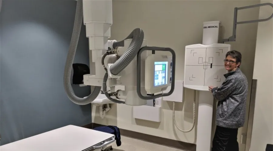
Radiographic Imaging (“X-Rays”)
Radiography uses ionizing radiation (x-rays) to image any part of the human anatomy from head to toe. This technique is well-suited to the skeleton.
Facilities, Technical Capabilities, and Limitations
- There are five radiography (x-ray) units available for human research.
- All units produce high-quality digital images.
- Images are viewable and archived on the Enterprise-wide McKesson PACS; CDs/DVDs can be created as needed.
- Radiology reports are viewable through SCM/AEHR and can be printed as needed.
| Unit | Location | Type | Table Weight Limits (kg) |
|---|---|---|---|
| 2 | Second Floor, Chandler Hospital, Pavilion H | Siemens Luminos Agile Max | 275 |
| 1 | Second Floor, Chandler Hospital, Pavilion H | Siemens Luminos dRF Max | 300 |
| 4 | First Floor KY Clinic (KYC) x-ray room | Del Medical OTC 15 | 360 |
| 1 | First Floor KY Clinic (KYC) x-ray room | Del Medical OTC 18 | 360 |
| 1 | First Floor KY Clinic (KYC) | Dexa/BMD - Hologic Discovery A | 200 |
Hours of Operation
- Monday – Friday, 7:30 a.m. – 4:30 p.m.
Hours of Operation
- Monday – Friday, 7:30 a.m. – 4:30 p.m.
Personnel Resources
- American Board of Radiology-certified, subspecialized Radiology Physicians (MDs).
- American Board of Radiology-certified Medical Physicist (PhD).
- American Registry of Radiologic Technologists (ARRT)-certified Radiology Technologists.
Personnel Resources
- American Board of Radiology-certified, subspecialized Radiology Physicians (MDs).
- American Board of Radiology-certified Medical Physicist (PhD).
- American Registry of Radiologic Technologists (ARRT)-certified Radiology Technologists.
Costs
- Please discuss project and all applicable costs with Radiology Senior Research Coordinator prior to IRB and/or grant submission or renewal.
- Radiology costs include the technologist’s time to prepare and image the patient/human research subject according to protocol, process the image data, and archive the images to a CD/DVD if required by the investigator or study sponsor.
- Supply costs might be incurred, e.g., CDs, DVDs
- Professional services by radiology physicians and/or medical physicists are negotiable.