Diffusion Weighted Magnetic Resonance Imaging (MRI) in Stroke
Blood and Clot Thrombectomy Registry and Collaboration (BACTRAC)
PI: Justin Fraser, MD; Co-I: Douglas Lukins
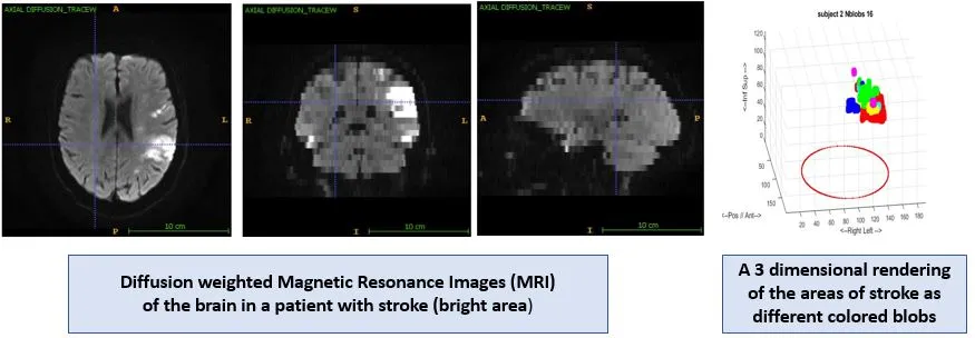
Douglas Lukins, MD, Assistant Professor of Radiology, is using diffusion-weighted Magnetic Resonance Imaging (MRI) of stroke patients to relate the site and volume of the stroke to the patient’s National Institute of Health (NIH) stroke scale. This study involves patients undergoing thrombectomy for stroke, during which blood samples are collected, capturing the thrombus along with arterial blood proximal and distal to it. The University of Kentucky labs in the Biomedical/Biological Science Research Building, (BSBSRB), have been an integral part of this research where clots are analyzed, to include gene analysis. This has been a joint clinical/basic science effort among multiple laboratories and the Neuro-Interventional Radiology service line, to include Douglas Lukins, MD and Abdulnasser Alhajeri, MD. As co-investigator Dr. Lukins conducts analysis on qualifying patient imaging including brain MRI and Computed Tomography Angiography (CTA). His analysis includes calculating infarct volumes and grading of hemorrhage, as well as grading of collaterals on the CTA. Collected information is being stored in a database for ongoing and future research for stroke patients.

