Ultrasound Imaging
Ultrasound uses high-frequency sound waves (not ionizing radiation, not x-rays) to create high-resolution two-dimensional ultrasound images of structures within the body. Ultrasound may be conducted safely in most volunteers, children, and pregnant women and may be repeated without risk.
Facilities, Technical Capabilities, and Limitations
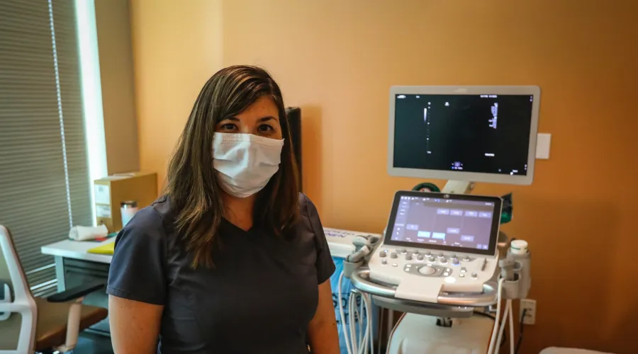
- Multiple diagnostic ultrasound units equipped with different frequency, linear-array, and phased-array transducers are available for a variety of human research applications.
- All units produce high-quality digital images.
- Images are viewable and archived on the Enterprise-wide McKesson PACS; images can be transcribed to CDs/DVDs as needed. Other methods to transfer electronic images are available.
- Radiology reports are viewable through SCM/AEHR and can be printed as needed.
Facilities, Technical Capabilities, and Limitations
- Multiple diagnostic ultrasound units equipped with different frequency, linear-array, and phased-array transducers are available for a variety of human research applications.
- All units produce high-quality digital images.
- Images are viewable and archived on the Enterprise-wide McKesson PACS; images can be transcribed to CDs/DVDs as needed. Other methods to transfer electronic images are available.
- Radiology reports are viewable through SCM/AEHR and can be printed as needed.
| Amount | Location | Equipment |
|---|---|---|
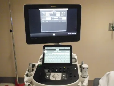
| 1 | Pavilion A | Philips iU22 |
| 5 | Pavilion A | Philips EPIQ |
| 2 | Pavilion A | Philips Affiniti |
| 1 | Pavilion H | Philips iU22 |
| 1 | Pavilion H | Philips CX50 |
Hours of Operation
- Monday - Friday, 7:30 a.m. - 5:00 pm
- Other days/times by arrangements
Hours of Operation
- Monday - Friday, 7:30 a.m. - 5:00 pm
- Other days/times by arrangements
Personnel Resources
- American Board of Radiology-certified, subspecialized Radiology Physicians (MDs).
- American Board of Radiology-certified Medical Physicist (PhD).
- American Registry for Diagnostic Medical Sonographers (ARDMS) and/or American Registry of Radiologic Technologists (ARRT)- certified, subspecialized Sonographers
Costs
- Please discuss project and all applicable costs with Radiology Senior Research Coordinator prior to IRB and/or grant submission or renewal.
- Radiology costs include the sonographer's time to prepare and image the patient/human research subject according to protocol, process the image data, and archive the images to PACS.
- Other archive media as required by the investigator or study sponsor; to be completed by Image Records.
- Professional services by radiology physicians and/or medical physicists are negotiable.