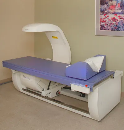
Dual Energy X-ray Absorptiometry (DXA)/Bone Mineral Density (BMD) Imaging
A DXA scanner uses low-level x-rays to measure bone mineral density (BMD) of the lumbar spine, hip, and/or forearm, typically for the diagnosis and monitoring of osteoporosis. A DXA scanner can measure whole-body tissue composition.
| Unit | Location | Type | Table Weight Limit (kg) |
|---|---|---|---|
| 1 | First Floor, Kentucky Clinic (KYC) | Hologic Horizon | 200 |
Facilities, Technical Capabilities, and Limitations
- In radiology, there is one DEXA scanner available for human research.
- The scanner produces high-quality digital images.
- Images are viewable and archived on the Enterprise-wide McKesson PACS; CDs/DVDs can be created as needed.
- Radiology reports are viewable through SCM/AEHR and can be printed as needed.
Hours of Operation
- Monday – Friday, 8 a.m. – 2 p.m.
Hours of Operation
- Monday – Friday, 8 a.m. – 2 p.m.
Personnel Resources
- American Board of Radiology and/or American Board of Nuclear Medicine-certified, subspecialized Radiology Physicians (MDs).
- American Board of Radiology-certified Medical Physicist (PhD).
- American Registry of Radiologic Technologists (ARRT)-certified Radiology Technologists with subspecialty certification in Bone Densitometry.
Costs
- Please discuss project and all applicable costs with Radiology Senior Research Coordinator prior to IRB and/or grant submission or renewal.
- Radiology costs include the technologist’s time to prepare and image the patient/human research subject according to protocol, process the image data, and archive the images to a CD/DVD if required by the investigator or study sponsor.
- Supply costs might be incurred, e.g., CDs/DVDs.
- Professional services by radiology physicians and/or medical physicists are negotiable.