Magnetic Resonance Imaging (MRI)
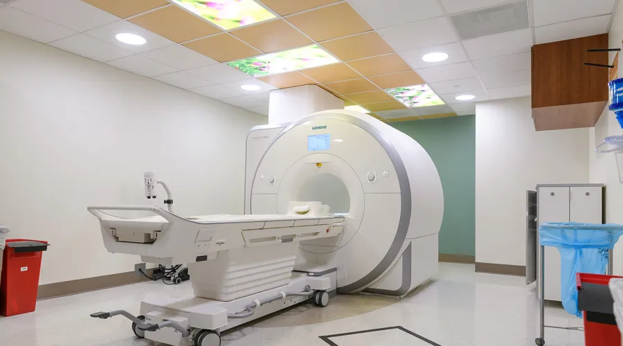
MRI uses strong magnetic fields and radiowaves, not x-rays, to create high-contrast three-dimensional tomographic images (“slices”) of any part of the human anatomy. Intravenous gadolinium-based contrast media may be given to enhance normal anatomy and delineate pathology. MRI may be conducted safely in most volunteers, including children, and pregnant women and may be repeated without risk. Imaging in patients with implanted devices (e.g., pacemaker) is generally contraindicated.
Facilities, Technical Capabilities, and Limitations
- There are six MR scanners available for human research.
- All scanners are high-field (1.5T or 3.0T), superconducting systems.
- All units produce high-quality digital images.
- Images are viewable and archived on the Enterprise-wide McKesson PACS; CDs/DVDs can be created as needed.
- Radiology reports are viewable through SCM/AEHR and can be printed as needed.
| Unit | Location | Type | Field Strength (T) | Special Clinical Applications | Table Weight Limit (kg) | Bore Size (cm) |
|---|---|---|---|---|---|---|
| 1 | Ground Floor, Pavilion A | Siemens Aera | 1.5 | Pediatrics (sedation) | 250 | 70 |
| 2 | Ground Floor, Pavilion A | Siemens Aera | 1.5 | Abdominal | 250 | 70 |
| 3 | Ground Floor, Pavilion A | Siemens Skyra | 3 | Neurological/ Musculoskeletal | 250 | 70 |
| 4 | Ground Floor, Pavilion G | Siemens Aera | 1.5 | Cardiac | 250 | 70 |
| 5 | Ground Floor, Pavilion G | Siemens Verio | 3 | Neurological | 250 | 70 |
| 6 | First Floor, Kentucky Clinic (KYC) | Siemens Aera | 1.5 | Breast | 250 | 70 |
Hours of Operation
- Monday – Friday, 7:30 a.m. – 7:00 p.m.
- Other days/times by arrangement.
Hours of Operation
- Monday – Friday, 7:30 a.m. – 7:00 p.m.
- Other days/times by arrangement.
Personnel Resources
- American Board of Radiology-certified, subspecialized Radiology Physicians (MDs).
- American Board of Medical Physics-certified Medical Physicist (PhD).
- American Registry of Radiologic Technologists (ARRT)-certified Radiology Technologists with subspecialty certification in MRI.
Costs
- Please discuss project and all applicable costs with Radiology Senior Research Coordinator prior to IRB and/or grant submission or renewal.
- Radiology costs include the technologist’s time to prepare and image the patient/human research subject according to protocol, process the image data, and archive the images to a CD/DVD if required by the investigator or study sponsor.
- Supply costs might be incurred, e.g., contrast media.
- Professional services by radiology physicians and/or medical physicists are negotiable.