Nuclear Medicine and Molecular Imaging
Nuclear Medicine uses gamma rays (a form of ionizing radiation similar to x-rays) emitted from a wide variety of radiopharmaceuticals to create images of the entire body from head to toe. The biodistribution pattern reflects normal physiology or underlying pathophysiology. The radiopharmaceuticals may be administered intravenously, orally, or by other routes.
Facilities, Technical Capabilities, and Limitations
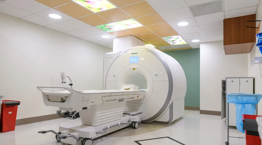
- There are three gamma camera units available for human research.
- All are capable of performing Single Photon Emission Computed Tomography (SPECT) (“slices”).
- All units produce high-quality digital images.
- There is one co-located thyroid probe available for human research.
- Images are viewable and can be analyzed on MIRADA, Medical XD, Siemens MMWP, DATAQUANT and McKesson PACS.
- Images can be archived on McKesson PACS and/or on other archive media such as CD/DVD.
- Radiology reports are viewable through SCM/AEHR and can be printed as needed.
Facilities, Technical Capabilities, and Limitations
- There are three gamma camera units available for human research.
- All are capable of performing Single Photon Emission Computed Tomography (SPECT) (“slices”).
- All units produce high-quality digital images.
- There is one co-located thyroid probe available for human research.
- Images are viewable and can be analyzed on MIRADA, Medical XD, Siemens MMWP, DATAQUANT and McKesson PACS.
- Images can be archived on McKesson PACS and/or on other archive media such as CD/DVD.
- Radiology reports are viewable through SCM/AEHR and can be printed as needed.
| Units | Location | Type | Table Weight Limit (kg) |
|---|---|---|---|
| 1 | Ground Floor, Chandler Hospital, Pavilion H | Siemens Symbia S | 227 |
| 2 | Ground Floor, Chandler Hospital, Pavilion H | Siemens S, (SPECT) | 200 |
| 3 | Ground Floor, Chandler Hospital, Pavilion H | Siemens Intevo Bold, (SPECT/CT) | 227 |
Hours of Operation
- Monday – Friday, 7:30 a.m. – 4:30 p.m.
- Other days/times by arrangement.
Hours of Operation
- Monday – Friday, 7:30 a.m. – 4:30 p.m.
- Other days/times by arrangement.
Personnel Resources
- American Board of Radiology and/or American Board of Nuclear Medicine-certified, subspecialized Radiology Physicians (MDs) who are Authorized Users of Radioactive Materials for Human Use.
- At least one of the Department of Radiology’s Authorized Users of Radioactive Materials must be named as a Co-Investigator.
- American Board of Radiology-certified Medical Physicist (PhD).
- Nuclear Medicine Technology Certification Board (NMTCB)-certified Nuclear Medicine Technologists and/or American Registry of Radiologic Technologists (ARRT (N))
Costs
- Please discuss project and all applicable costs with Radiology Senior Research Coordinator prior to IRB and/or grant submission or renewal.
- Radiology costs include the technologist’s time to prepare and image the patient/human research subject according to protocol, process the image data, and archive the images to a CD/DVD if required by the investigator or study sponsor.
- Supply costs might be incurred, e.g., radiopharmaceuticals.
- Professional services by radiology physicians and/or medical physicists are negotiable.