Hyde Lecture: “Imaging and Navigation in Complex Cardiovascular Pathologies”
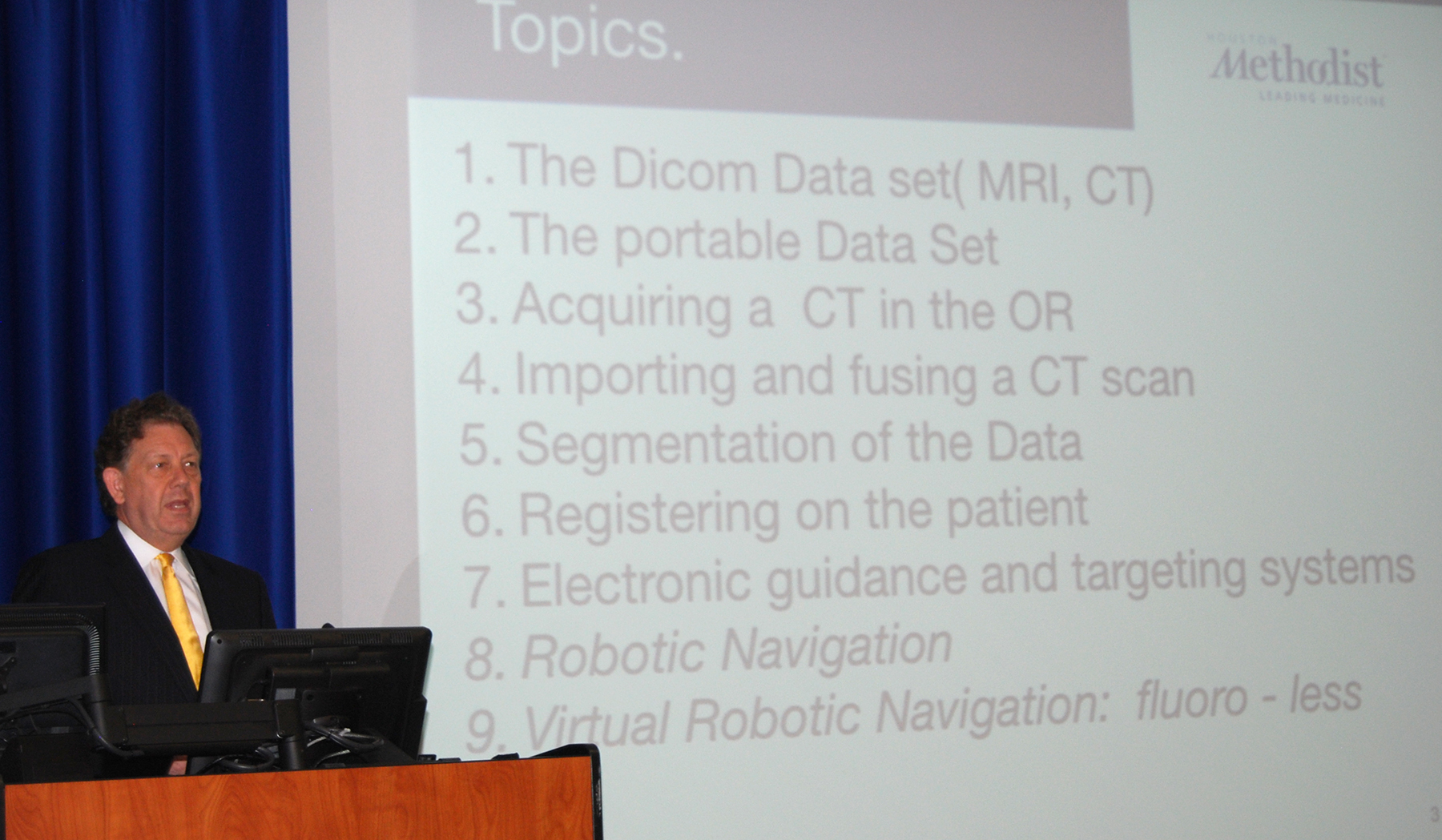
The “Holy Grail” for vascular surgery technology in the next five to six years will be the fusion of three-dimensional CT images to robotic navigation in a hybrid operating room, said Dr. Alan B. Lumsden, chair of the Houston Methodist Department of Cardiovascular Surgery, who delivered the inaugural lecture for the Gordon L. Hyde Lectureship in Vascular Surgery.
[Insert link to Dr. Lumsden’s Powerpoint Presentation]
Lumsden holds the Walter W. Fondren III Distinguished Endowed Chair with the DeBakey Heart and Vascular Center at Houston Methodist. He is one of the key national investigators in NIH-funded research for the development of “novel minimally invasive methods of surgery.”
The title of his lecture, “Imaging and Navigation in Complex Cardiovascular Pathologies,” discussed the next generation of advanced 3D imaging technology and its applications in complex cardiac, vascular, and thoracic surgery, among other fields.
Though his formal training is as a vascular surgeon, he described himself as “an imaging guy.” And he told all the active surgeons, and particularly the residents and fellows in the lecture hall that they would be the same. “You’re all going to be imaging people. There’s no way to avoid it. Practitioners are going to be relying heavily on imaging” for greater accuracy and greater control, Lumsden said.
Elaborating further, Lumsden described how surgeons will not only use 3D CT imaging to map out procedures in advance of engaging their patient in the operating room, they will take that image and virtually “lay it on top of the patient and interact directly with the CT image.”
But even more than just using a single CT image, researchers at Houston Methodist are experimenting with the fusion of existing images with cone beam CT Images created in the hybrid operating room prior to the procedure. Such image fusion would present surgeons with a highly accurate three dimensional display of an individual’s cardiovascular system and highlight the areas requiring surgical intervention or repair.
From that information, Lumsden said, surgeons can navigate minimally invasive instruments along the most direct path to perform any number of complex procedures such as translumbar embolization, aortic aneurysm surgery, lung nodule localization and biopsy, pulmonary embolism, or even in transcatheter aortic valve replacement (TAVR) or implantation (TAVI) procedures.
This form of imaging fusion is not new, Lumsden continued. The techniques and technology are already being applied on a daily basis “in the neurosurgical world, the thoracic surgery world, and even in the TAVR world” where the need for minimally invasive procedures is most desirable for better outcomes.
What researchers are ultimately working toward is combining highly detailed imaging systems to robotic navigation, allowing surgeons to carefully guide needles and microsurgical tools directly where they need to go. He referred to that fusion of technologies as the “Holy Grail” of their research, but in this case, the goal is real and achieveable within the next decade.
