Tori Stromp Successfully Defends Her Dissertation
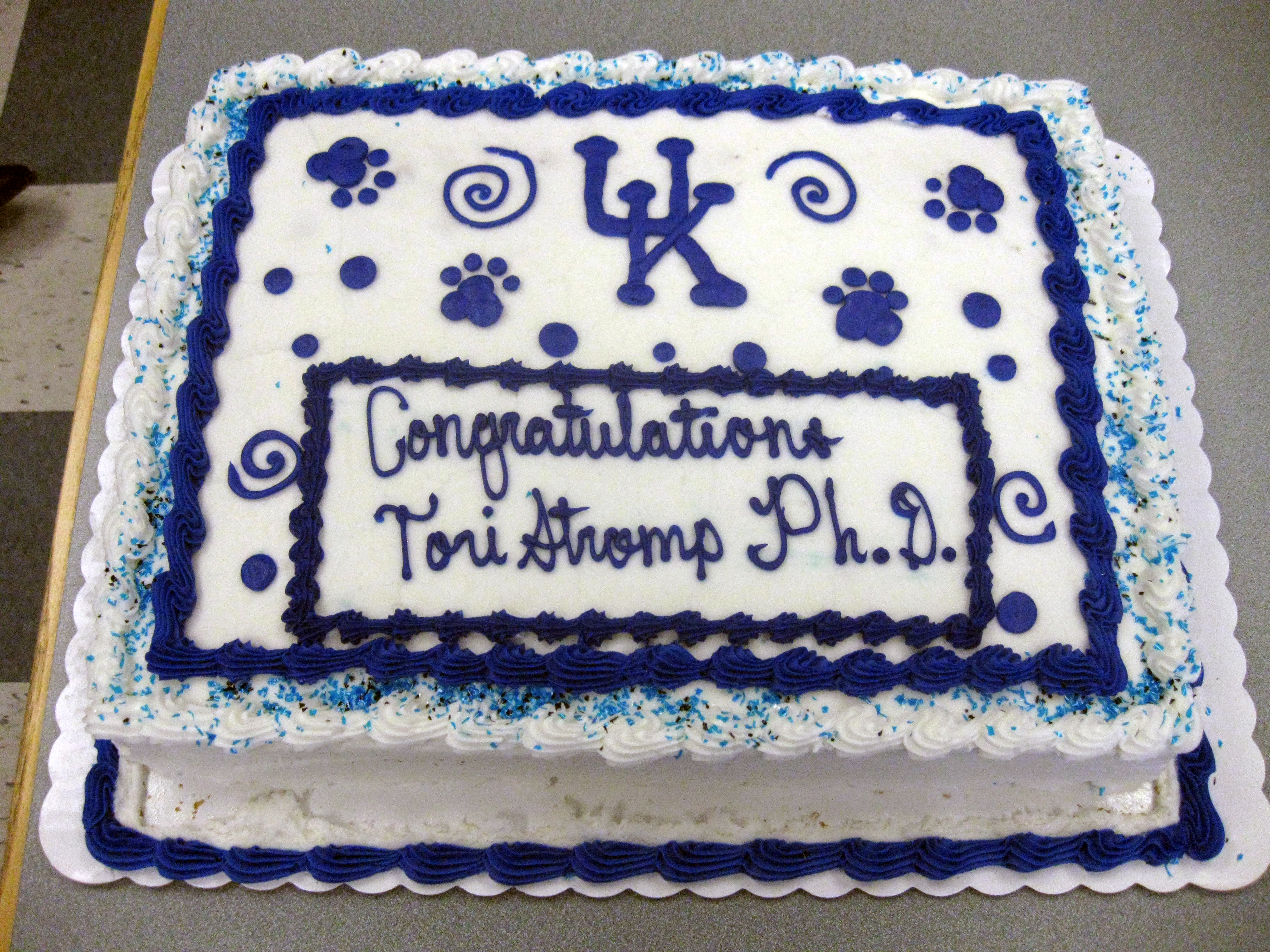
Congratulations Tori Stromp, PhD! Tori successfully defended her dissertation on November 28, 2016.
"Development and Application of Gadolinium Free Cardiac Magnetic Resonance Fibrosis Imaging for
Multi-Scale Study of Heart Failure in Patients with End Stage Renal Disease"
Abstract of Dissertation
Cardiac magnetic resonance (CMR) is a powerful tool to noninvasively image ventricular fibrosis. Late gadolinium enhancement (LGE) CMR identifies focal and, with T1 mapping, diffuse fibrosis. Despite prevalent cardiac fibrosis and heart failure, patients with end stage renal disease (ESRD) are excluded from LGE. Absence of a suitable diagnostic has limited the understanding of heart failure and obstructed development of therapies in the setting of ESRD. A quantitative, gadolinium free fibrosis detection method could overcome this critical barrier, propelling the advancement of diagnostic, monitoring, and therapy options. This project describes the development of a gadolinium-free CMR technique and application for cardiac fibrosis measurement in patients with ESRD.
Magnetization transfer (MT) occurs during standard cine balanced steady state free precession (bSSFP) CMR, where extracellular matrix protons exchange magnetization with water molecules. Extracellular water volume expansion, concomitant with fibrosis, reduces MT and subtly elevates signal intensity. Our technique, 2-pt bSSFP, extracts endogenous contrast sensitive to tissue fibrosis by obtaining pairs of high and low MT-weighted images and calculating normalized signal differences, denoted by ΔS/So
We tested 2-pt bSSFP in patients referred for CMR and found excellent agreement spatially with LGE and quantitatively with extracellular volume fraction. Diagnostic and clinical application of 2-pt bSSFP was comparable to LGE. We applied 2-pt bSSFP to patients with ESRD for multi scale comparison with correlates of fibrosis ranging from blood biomarkers to whole organ function. Patients with ESRD displayed hypertrophy with reduced contraction, but elevated ΔS/So and fibrosis. Some biomarkers correlated with both hypertrophy and fibrosis, highlighting the need to distinguish between hypertrophic and fibrotic remodeling. We monitored fibrosis over 1 year using 2-pt bSSFP in a cohort of patients with ESRD. ΔS/So and fibrotic burden increased substantially, despite minor changes in structure and function.
Collectively these studies validate and apply 2-pt bSSFP for gadolinium free fibrosis CMR in patients with ESRD. While ventricular structure and function are commensurate with progression toward heart failure, it is now possible to specifically describe global and focal patterns of cardiac fibrosis in ESRD, along with comparisons to blood biomarkers, which may lead to improved diagnostics and molecular treatment targets.
Doctoral Committee Members
Dr. Moriel Vandsburger Department of Physiology
Dr. Brian Delisle Department of Physiology
Dr. Brian Jackson Department of Physiology
Dr. Susan Smyth Department of Physiology
Dr. Gregory Graf Department of Pharmaceutical Sciences
Outside Examiner
Dr. Misung Jo Department of Obstetrics and Gynecology
Acknowledgements
It takes a village to raise a graduate student. I owe immeasurable gratitude to the following individuals, among many others, for their impact on my training and this project.
I am truly grateful for the guidance of my primary mentor and Dissertation Chair, Dr. Moriel Vandsburger. His unmatched intelligence, unwavering assurance, and unending positivity have driven my eternal pursuit of inquiry and success with an ingrained, genuine optimism. I aspire to someday achieve the caliber of scientist and mentor that I was so fortunate to encounter.
I gratefully acknowledge my Dissertation Committee—Dr. Brian Delisle (Co-Chair), Dr. Brian Jackson, Dr. Susan Smyth, and Dr. Gregory Graf—for their constant challenge and direction, which have improved me as a scientist through this process. I thank Dr. Misung Jo for serving as the external examiner for my dissertation defense.
Dr. Steve Leung deserves the utmost appreciation for his detailed training. He taught me the balance of technical skill, troubleshooting, and patient rapport which merely scratches the surface of his professional and scientific prowess. Many thanks to Dr. Vincent Sorrell for his mentorship and assistance with our protocols, and the cardiac MRI technicians—Becca Egli, Joe Jenkins, John Green and Karsten Colwell —for their assistance in image acquisition. I am grateful for the contributions of Tyler Spear, Becca Kidney, Kristin Andres, and Josh Kaine, who were game-changers at pivotal points throughout this work. Each has truly improved the course of this research. It was a privilege to work with these young stars who are destined to impact the future of science and medicine.
My partner, Carl Flotka—I cannot express the depth to which your support has and will continue to affect my life. I could have done this without you, but I am sure glad I didn’t have to.
Finally, I extend my deepest gratitude to the dedicated participants of this research. The altruism of these volunteers exhibits their commitment to the progress of science and medicine. While many will never witness or benefit from the improvements we aim to make in clinical care, their time, data, and insight will forever impact our understanding of disease and drive advances in diagnostic and treatment options. They are, after all, why I became a scientist.
