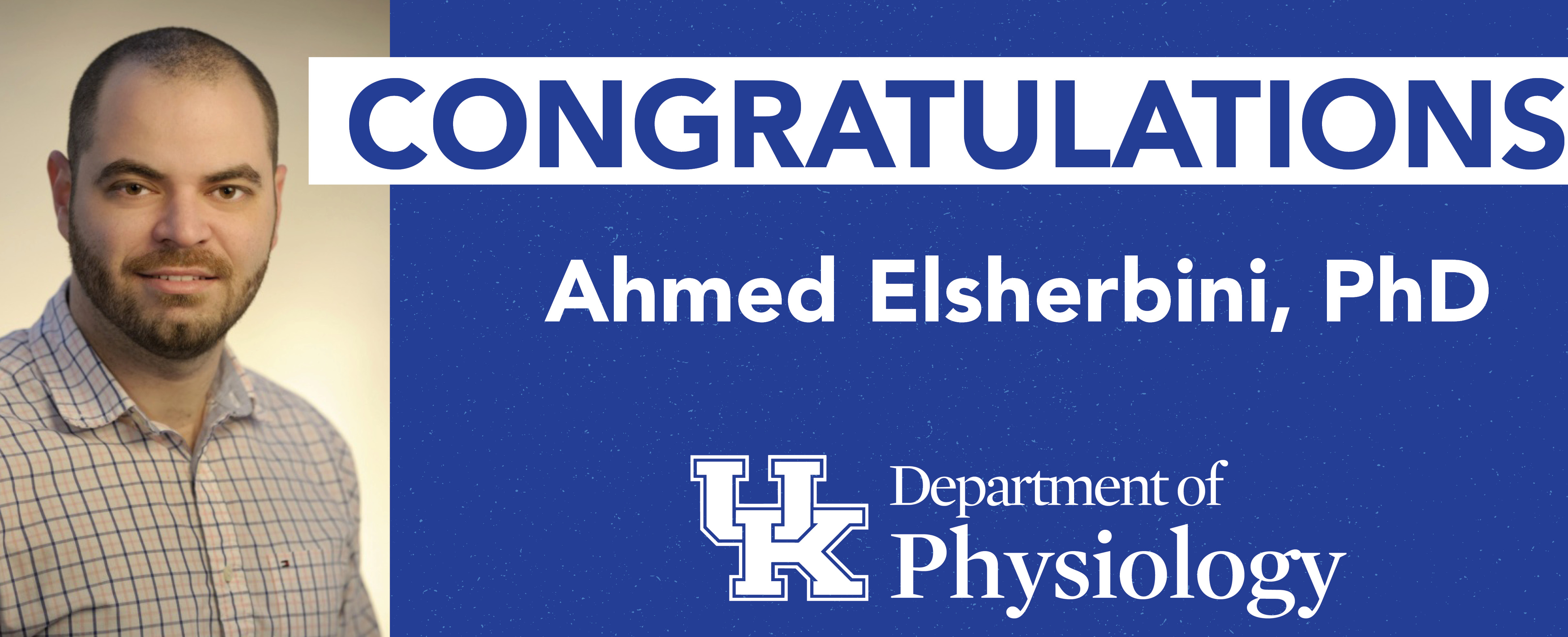
Congratulations Ahmed Elsherbini, PhD
On Wednesday, November 11, 2020 Ahmed Elsherbini successfully defended his dissertation and earned his doctoral degree. Congratulations Dr. Elsherbini!
"Ceramide-enriched extracellular vesicles: A role in enhancing Amyloid-beta neurotoxicity and mitochondrial damage in Alzheimer’s disease"
Doctoral Committee Members
Dr. Erhard Bieberich, Department of Physiology, Mentor
Dr. Mariana Nikolova-Karakashian, Department of Physiology
Dr. Steven Estus, Department of Physiology
Dr. John McCarthy, Department of Physiology
Dr. Vivek Rangnekar, Department of Toxicology
Dr. Penni Black, Outside Examiner, Department of Pharmaceutical Science
Abstract
Alzheimer’s disease (AD) is an age-dependent, progressive, neurodegenerative disorder that is characterized clinically by the impairment of cognitive functions concomitant with behavioral and personality changes. AD is associated with distinct pathological hallmarks, namely, intracellular neurofibrillary tangles comprised of hyperphosphorylated tau protein, extracellular amyloid beta (Aβ) plaques, and marked brain atrophy. Besides their main rule as the core component of amyloid plaques, oligomeric Aβ have been shown to be neurotoxic. The exact mechanism of Aβ neurotoxicity is yet to be elucidated. Recently, a pathogenic function of small extracellular vesicles- also known as exosomes- has been proposed, suggesting that exosomes can transfer pathogens between cells. One such pathogen that exploits this pathway is Aβ in Alzheimer’s disease, however, it is not known yet whether this Aβ/exosomes association would affect the neuronal toxicity of Aβ. Exosomes are nano-sized lipid vesicles that are formed by inward budding of late endosomes to form multi vesicular bodies (MVB) which fuse to the plasma membrane and release exosomes to the extracellular space. Exosomes serve as a means of intercellular communication due to their ability in carrying cargoes including microRNA (miRNA), messenger RNA (mRNA), proteins, and other biomolecules. There are several established pathways for exosomes biogenesis, one of which is triggered by the sphingolipid ceramide. Ceramide is a key molecule in sphingolipids metabolism and it is involved in several cellular processes such as proliferation, senescence and apoptosis. It has also been reported that ceramide levels are elevated in AD patients brain specimens. Exploiting the fact that exosomes can cross the blood brain barrier we therefore used serum derived exosomes to study the biophysical and biochemical characteristics of Alzheimer’s disease mouse model (5xFAD) and AD patients’ exosomes compared to wild type and healthy individuals. We found that serum from 5xFAD mice and AD patients contain a subpopulation of astrocyte-derived exosomes that are enriched with ceramide, particularly C16:0, C18:0, C20:0, 22:0, C24:0, and C24:1 ceramide species. This subpopulation (termed astrosomes) were shown to associated with Aβ and they are prone to aggregation as confirmed by nanoparticle tracking and cluster analyses. To study the functional characteristics of these Aβ-associated astrosomes, we used Neuro-2a (N2a) cells, human iPS cell-derived neurons, and mouse primary cultured neurons as in vitro tissue culture models. When taken up by neurons, Aβ-associated astrosomes were specifically transported to mitochondria where they induced mitochondria clustering, evident by elevation of expression of the fission protein dynamin related protein1 (Drp1). Aβ-associated astrosomes, but not wild type or healthy control human exosomes, mediated binding of Aβ to voltage-dependent anion channel 1 (VDAC1), a gate keeper protein in the outer mitochondrial membrane that is involved in regulating passage of metabolites, nucleotides, and ions; it plays a crucial role in regulating apoptosis. This Aβ/VDAC1 interaction leds to caspase activation and subsequently apoptosis. Interestingly, removing the ceramide-enriched astrosomes from the exosome pool using lipid-mediated affinity chromatography (LIMAC) mitigated that toxic effect on neurons. These results were replicated using brain derived exosomes. To investigate the in vivo significance of our in vitro results, we stereotaxically injected wild type mice (two weeks old) with 5xFAD or wild type brain derived exosomes (nine months old). We found that within two days, the injected exosomes were specifically taken up by neurons and transported to mitochondria. Consistent with our in vitro data using Aβ-associated astrosomes, the exosomes isolated from 5xFAD brain, but not those from wild type brain, induced complex formation of Aβ with VDAC1 and activation of caspase 3. To test that our observations hold true in physiological conditions, we generated a novel astrosome reporter mouse model. This was accomplished by crossing of Aldh1l1-Cre/ ERT with floxed CD63-GFP and 5xFAD mice (5XFAD xAldh1l1-Cre/ERTxCD63-GFPfl/fl) which allows us to track astrosome uptake and their subsequent effects. As seen with the injected exosomes, we found that endogenous GFP-labeled astrosomes are taken up by neurons where they shuttle Aβ and induce mitotoxicity. In conclusion, our data show that association of Aβ to astrasomes in critical for Aβ neurotoxicity. Therefore, we discovered a novel mechanism by which Aβ induces AD neuropathology as well as potential pharmacological target.
Acknowledgements
First and foremost, I would like to extend gratitude to Dr. Erhard Bieberich for all his guidance, patience and motivation throughout the past few years. Thank you for giving me enough autonomy while continuously mentoring the scientific method and experimental rigor. Thank you for taking a chance on me, pushing me out of my comfort zone, believing in me when I was in doubt, and for challenging me to become a better scientist. To my committee members, Dr. Steve Estus, Dr. Mariana Nikolova-Karakashian, Dr. John McCarthy and Dr. Vivek Rangnekar I’m grateful for all your time, patience and advices along the way. Your constructive comments and questions contributed positively to my scientific growth and helped accomplishing the work in its final form. I would also like to thank the whole PGY community, student, lecturers, staff for making it as easy as possible to navigate through this journey. I am forever indebted to my parents for all their sacrifices and support since I could remember. None of what I have done would have been possible without your prayers, support and unconditional love. You both are an example of how ideal parents should be, and I wish I could be as good as you are. My sisters, Amira and Nihal, and my bother Mido I do appreciate your continuous encouragement and help. Whether it was a phone call, text or a facebook comment; your communication has always been a fuel for me to keep moving and never give up, love you all. I was lucky to have worked alongside a long list of lab mates with whom I shared several moments of joys, hippieness and a few tears. Thank you all for the thought-provoking discussions, technical help and for providing a good working atmosphere. Finally, and most importantly, I have to admit the last few years would be impossible without the love, support, and understanding of my wife, Sara, and my daughters Farida and Layla. I am at loss for words and can’t even begin to describe how lucky I am to have you as my friend, wife, soulmate, and sometimes mother. I realize all the sacrifice you have been doing throughout the years starting from coming all the way to U.S.A, putting up with late days at work and me working on weekends and much more. Thank you for being my inspiration and backbone, and for allowing me to pursue the career I chose, despite being a risky move with unknown outcomes. My daughters, you’re the breath of fresh air each day. Whenever I’m coming back home from work depressed or overwhelmed, it all goes away when I hug you both, I can’t wait to see both of you leading a great successful life you deserve.
