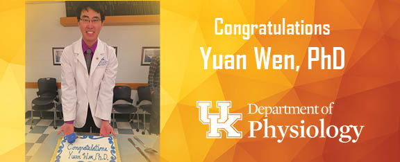
Congratulations Yuan Wen, PhD
On December 12, 2017 Yuan Wen successfully defended his dissertation and earned his PhD. With the basic science research portion of his MD/ Ph.D. program complete, Dr. Wen will now return to the clinical side to continue working towards his MD. Congratulations Dr. Wen!
β-CATENIN REGULATION OF ADULT SKELETAL MUSCLE PLASTICITY
Doctoral Committee Members
Dr. John McCarthy, Department of Physiology
Dr. Steve Estus, Department of Physiology
Dr. Charlotte Peterson, Department of Rehabilitation Sciences
Dr. Tim McClintock, Department of Physiology
Dr. Ken Campbell, Department of Physiology
Dr. Elizabeth Debski, Department of Biology (Outside Examiner)
Abstract
Skeletal muscle is estimated to be more than 40% of total body mass in a healthy adult, almost half of the body’s total protein and amino acid storage. Muscle mass deficit impacts total body metabolism and homeostasis, and muscle loss is the most significant predictor of bone loss. Numerous forms of disease and injury can lead to detrimental losses of muscle mass. Malignancies, congestive heart failure, chronic obstructive pulmonary disease, cystic fibrosis, rheumatoid arthritis, Alzheimer’s disease, infections, and many other forms of chronic pathologies all lead to symptoms of cachexia with varying rates of prevalence. Significant loss of skeletal muscle mass dramatically reduces quality of life for patients with cancer, pathological aging, neurological injuries, protein-energy wasting, and critical illnesses. The association of skeletal muscle loss with poor clinical outcomes and low quality of life is strong, consistent, and dose-dependent. Muscle loss due to aging alone is a major burden on total US healthcare costs, while overall muscle atrophy and weakness can increase total healthcare costs per patient by more than 2-fold. Regardless of the cause of disease, muscle atrophy as a symptom is costly both for the patients and the economy. Even after successful treatment of disease, there is currently no effective method for improving patient muscle mass and strength to a level conducive for physical therapy and rehabilitation. A better understanding of the mechanisms of muscle growth will facilitate the development of bridge therapies that can significantly speed up patient recovery.
One of the most commonly utilized methods in studying skeletal muscle and quantifying myofiber hypertrophy is the use of image analysis with immunofluorescence microscopy. Skeletal muscle samples are typically cut in transverse or cross sections, and antibodies against sarcolemmal or basal lamina proteins are used to label the cell boundaries. The quantification of hundreds to thousands of cells per sample are accomplished either manually or semi-automatically using generalized pathology software, and such approaches become exceedingly tedious. In the first study, we developed a robust, fully automated software that is dedicated to skeletal muscle immunohistological image analysis. We have made this software freely available to muscle biologists to alleviate the burden of routine image analyses. To date, more than 50 technicians, students, postdoctoral fellows, faculty members, and others have requested our software.
Using the automated software, I was able to quickly quantify the effects of β-Catenin knockout on myofiber hypertrophy in the second study. In this study, we tested the hypothesis that myofiber hypertrophy is mediated by β-Catenin activation of c-myc and ribosome biogenesis. Previous studies have demonstrated the importance of ribosome biogenesis in cardiac muscle hypertrophy. Recent evidence in both mice and human suggest a close association between ribosome biogenesis and skeletal muscle hypertrophy. Using an inducible skeletal myofiber specific genetic knockout mouse model, we confirmed the requirement of β-Catenin for increased levels of c-myc and Pre-47S transcripts as well as myofiber hypertrophy.
Finally, we measured the effects of unilateral myofiber knockout of β-Catenin on resident muscle stem cells, or satellite cells. Junctional complexes on the surface of satellite cells in the niche between myofiber sarcolemma and the basal lamina are important for proper control of satellite quiescence. The muscle specific cadherin, Mcad, forms complexes with β-Catenin at the interface between myofiber and satellite cell membranes. Knockout of cadherins in satellite cells induces transition out of quiescence, and this effect is mediated through β-Catenin in the satellite cells. Although significant focus has been focused on the mechanisms within satellite cells, our study using a myofiber unilateral knockout of β-Catenin provide novel insights into the nature of junctional contact mediated quiescence in the myofiber-satellite cell niche. We show that losing β-Catenin in myofibers allow earlier activation and proliferation of satellite cells, which significantly enhances muscle regeneration. Our model may shed more light on the intricacies of satellite cell priming and transition to an activated state, with implications for regenerative medicine.
Acknowledgements
First and foremost, I would like to thank Dr. John McCarthy for all of his guidance, support, and motivation over the past few years. I couldn’t have asked for a better advisor and mentor. Not only has he shaped the way I ask questions and view scientific problems, but he has also been instrumental in helping me define my career goals. I wouldn’t be doing what I am and going where I plan without his support and inspiration.
Much of the work I’ve been able to accomplish in my dissertation is also thanks to the vital support and resources provided by Dr. Charlotte Peterson. Like, John, Char has been extremely supportive and together, they have enabled me to pursue a direction of research involving automation. Of course, this direction, especially the computation portion, wouldn’t have been possible without the mentorship and encouragement of Dr. Kenneth Campbell.
I would also like to thank Drs. Tim McClintock and Steve Estus for their insights and guidance over the past few years. I appreciate their advice on all the changes in directions I’ve had to take since the beginning of my graduate studies. Additionally, I would like to thank Dr. Susan Smyth, Therese Stearns, and the rest of the MD/PhD Program for their care over the past few years and also for their continued support in the years to come.
I am truly fortunate to have learned from and continue to work with such wonderful mentors, but I must also thank everyone who has made scientific research a joy for me in the last few years. Dr. Alexander Alimov has worked tirelessly to ensure everything is running and in tiptop shape so that everyone can continue to make progress. Drs. Ivan Vechetti and Kevin Murach have taught me how to approach team science and collaborate closely with others to design experiments and test hypotheses.
I also want to thank Dr. Cory Dungan for his clutch save during the final stretches of the β-catenin experiments. Drs. Vandre Casagrande Figueiredo, Chris Fry, Tyler Kirby, Thomas Chaillou, Esther DupontVersteegden, Janna Jackson, Sarah White, Grace Walton, and Kate Kosmac have provided invaluable expertise, experience, insightful discussions, and moral support. It’s been wonderful working together and I look forward to our continued collaborations.
It goes without saying that I wouldn’t be here without my parents. They raised me, brought me to America, and gave me the chance at opportunities I would never see otherwise. I hope they are proud of my efforts.
Finally, the last few years would be impossible without the love, support, and understanding of my wife, Fei, my son, Ethan, and my daughter, Ava. Experiments often take up time on nights and weekends and have taken a considerable amount of my time away from my family, but it means the world to me that they continue to believe in their husband and father. I can’t thank Fei enough for the sacrifices she’s had to make in her own education and career in order to accommodate my training. I’d like to say I’m done with school, but I still have a couple years left.
