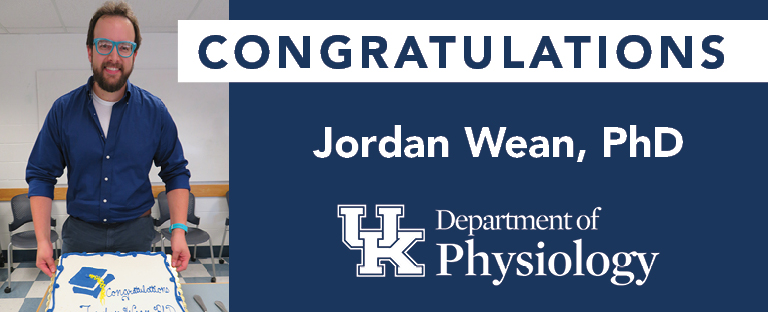
Congratulations Jordan Wean, PhD!
On Wednesday, July 28, 2021 Jordan Wean successfully defended his dissertation and earned his doctoral degree. Congratulations, Dr. Wean!
METABOLIC AND ELECTROPHYSIOLOGICAL EFFECTS OF FIBROBLAST GROWTH FACTOR 19 IN THE DORSAL VAGAL COMPLEX
Doctoral Committee Members
Dr. Bret Smith, Department of Neuroscience, Mentor
Dr. Olivier Thibault, Pharmacology & Nutritional Sciences
Dr. John McCarthy, Department of Physiology
Dr. Ken Campbell, Department of Physiology
Dr. Lisa Tannock, Department of Internal Medicine
Dr. Tadahide Izumi, Department of Toxicology and Cancer Biology, Outside Examiner
Abstract
The dorsal vagal complex (DVC) is an important homeostatic regulatory center located in the hindbrain that alters vagal parasympathetic activity in response to viscerosensory and humoral cues. Within the DVC, second-order sensory neurons in the nucleus tractus solitarius (NTS) integrate ascending vagal sensory input with descending regulatory inputs from higher brain areas and respond to circulating hormones and glucose. In turn, the NTS projects to the dorsal motor nucleus of the vagus (DMV) which is comprised of cholinergic motor neurons and regulates gastric motility, hepatic glucose production, and pancreatic hormone release among others. Fibroblast growth factor 19 (FGF19) is a protein hormone that produces antidiabetic effects when administered intracerebroventricularly in the forebrain. Lateral or third ventricle administration of FGF19 was shown to increase glucose tolerance, decrease body weight, and decrease food intake. However, no studies have been performed to understand the effects of FGF19 in the DVC. Neurons in the DVC express FGF receptors and regulate many of the processes that have been proposed to explain the antidiabetic effects of FGF19. Thus, this study was understand both the cellular effects of FGF19 in the DVC as well as effects on systemic glucose regulation. First, the FGF19 was applied to the hindbrain to understand its effects on blood glucose. Fourth ventricle administration of FGF19 produced no effect on blood glucose concentration in control mice, but induced a significant, peripheral muscarinic receptor-dependent decrease in systemic hyperglycemia for up to 12 hours in streptozotocin (STZ)-treated mice, a model of type 1 diabetes. Patch-clamp recordings from DMV neurons in vitro revealed that FGF19 application altered synaptic and intrinsic membrane properties of DMV neurons, with the balance of FGF19 effects being significantly modified by a recent history of systemic hyperglycemia. Since the previous data indicated that FGF19 alters firing in glutamatergic neurons upstream from the DMV, the next study was aimed at understanding the electrophysiological effects of FGF19 on local glutamatergic neurons in the DVC. Receptor expression studies indicated that two nuclei were the most likely source of glutamatergic input to the DMV: the NTS and the area postrema (AP). Glutamate photolysis studies indicated that FGF19 does indeed increase the activity of glutamatergic neurons in the AP and NTS that project to the DMV. This effect was only seen in hyperglycemic mice. Further study indicated that FGF19 produced mixed effects on the intrinsic excitability of NTS neurons but increased action-potential dependent glutamate release to the NTS in hyperglycemic mice. The source of this glutamate was confirmed to be the AP. Overall, the in vitro effect of FGF19 on DVC neuron excitability was complex. FGF19 produced mixed effects on the intrinsic excitability of some cells while substantially increasing glutamatergic transmission at multiple synapses in the DVC of hyperglycemic mice. In vivo, FGF19 in the hindbrain decreased blood glucose in diabetic mice, an effect that is consistent with its observed in vitro effects on glutamatergic transmission. These findings identify central parasympathetic circuitry as a novel target for FGF19 and suggest that FGF19 acting in the dorsal hindbrain can alter vagal output to produce its beneficial metabolic effects.
Acknowledgements
I would like to thank my mentor, Dr. Bret Smith for the advice and guidance he provided along the way. I thank my advisory committee members: Dr. Olivier Thibault, Dr. Lisa Tannock, Dr. Ken Campbell, and Dr. John McCarthy. They asked the tough questions and helped bring new perspectives to my research. I thank the current and past members of the Smith laboratory who helped me along the way. To point out a few members regarding their roles. Thank you to: Drs. Jeff and Carie Boychuk, Cory Butler, and Isabel Derera for teaching me electrophysiology. Also, I would like to thank Dr. Katalin Smith, who helped so much with my failed molecular biology experiments and who also provided endless tasty Hungarian food. I would like to acknowledge my family. I thank my parents, David and Pamela Wean who always inspired me to be curious, courageous, and conscientious. I thank my siblings, David and Chloe Wean for all the amazing and strange things they do. Finally, I cannot thank enough my wife Alyssa Wean, without whom, I would not be who I am and where I am today. She has been my best friend for as long as I can remember and an immense source of support, happiness, and advice though everything.
