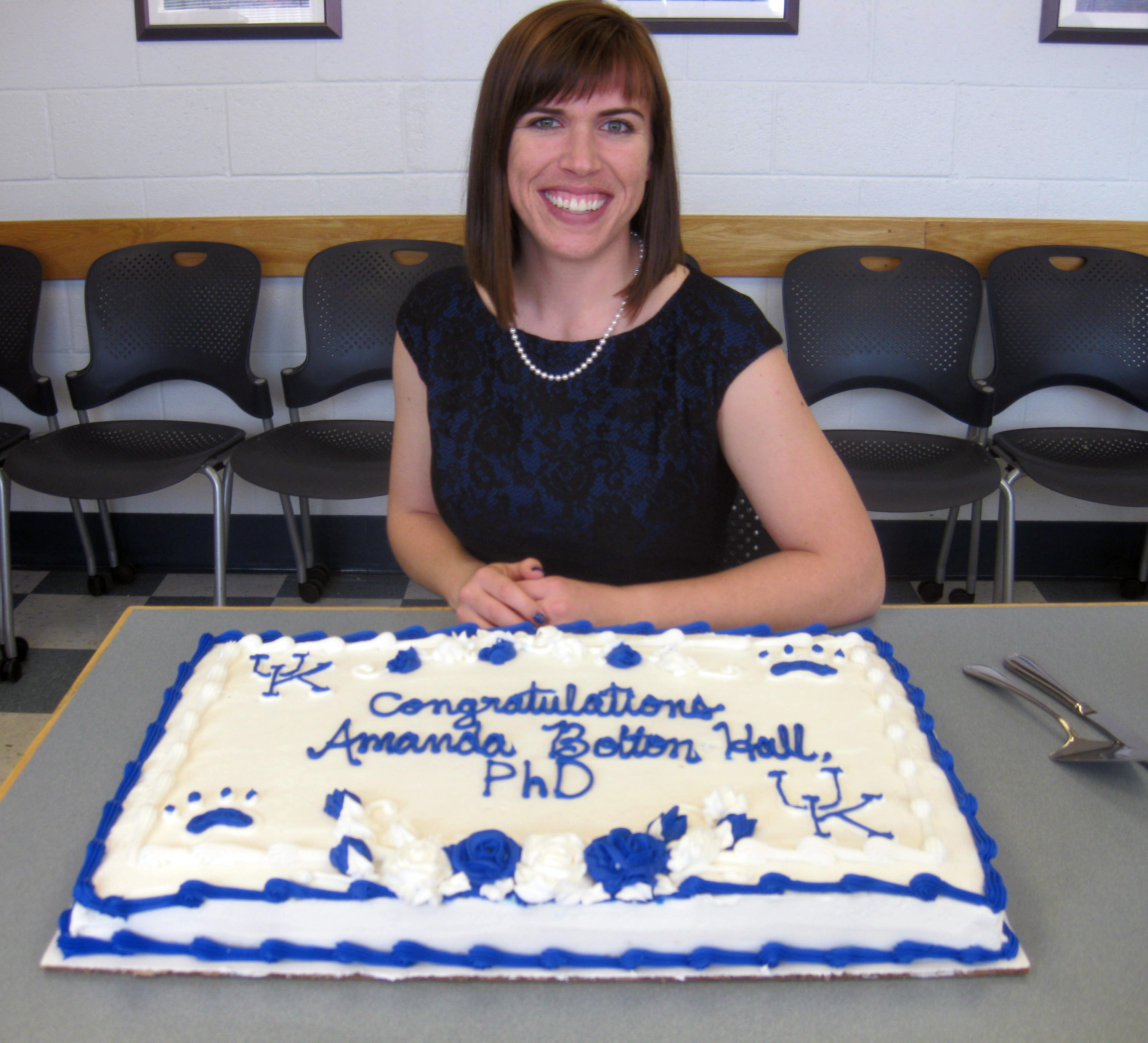
Congratulations Amanda Bolton Hall, Ph.D.
On April 20th, 2016, Amanda Bolton Hall successfully defended her dissertation.
“Histological and Behavioral Consequences of Repeated Mild Traumatic Brain Injury in Mice”
Abstract of Dissertation
The majority of the estimated three million traumatic brain injuries that occur each year are classified as “mild” and do not require surgical intervention. However, debilitating symptoms such as difficulties focusing on tasks, anxiety, depression, and visual deficits can persist chronically after a mild traumatic brain injury (TBI) even if an individual appears “fine”. These symptoms have been observed to worsen or be prolonged when an individual has suffered multiple mild TBIs. To test the hypothesis that increasing the amount of time between head injuries can reduce the histopathological and behavioral consequences of repeated mild TBI, a mouse model of closed head injury (CHI) was developed. A pneumatically controlled device with a silicone tip was used to deliver a diffuse, midline impact directly onto the mouse skull. A 2.0mm intended depth of injury caused a brief period of apnea and increased righting reflex response with minimal astrogliosis and axonal injury bilaterally in the entorhinal cortex, optic tract, and cerebellum. When five CHIs were repeated at 24h inter-injury intervals, astrogliosis was exacerbated acutely in the hippocampus and entorhinal cortex compared to a single mild TBI. Additionally, in the entorhinal cortex, hemorrhagic lesions developed along with increased neurodegeneration and microgliosis. Axonal injury was observed bilaterally in the white matter tracts of the cerebellum and brainstem. When the inter-injury interval was extended to 48h, the extent of inflammation and cell death was similar to that caused by a single CHI suggesting that, in our mouse model, extending the inter-injury interval from 24h to 48h reduced the acute effects of repeated head injuries. The behavioral consequences of repeated CHI at 24h or 48h inter-injury intervals were evaluated in a ten week longitudinal study followed by histological analyses. Five CHI repeated at 24h inter-injury intervals produced motor and cognitive deficits that persisted throughout the ten week study period. Based upon histological analyses, the acute inflammation, axonal injury, and cell death observed acutely in the entorhinal cortex had resolved by ten weeks after injury. However, axonal degeneration and gliosis were present in the optic tract, optic nerve, and corticospinal tract. Extending the inter-injury interval to 48h did not significantly reduce motor and cognitive deficits, nor did it protect against chronic microgliosis and neurodegeneration in the visual pathway. Together these data suggested that some white matter areas may be more susceptible to our model of repeated mild TBI causing persistent neuropathology and behavioral deficits which were not substantially reduced with a 48h inter-injury interval. In many forms of TBI, microgliosis persists chronically and is believed to contribute to the cascade of neurodegeneration. To test the hypothesis that post-traumatic microgliosis contributes to mild TBI-related neuropathology, mice deficient in the growth factor progranulin (Grn-/-) received repeated CHI and were compared to wildtype, C57Bl/6 mice. Penetrating head injury was previously reported to amplify the acute microglial response in Grn-/- mice. In our studies, repeated CHI induced an increased microglial response in Grn-/- mice compared to C57Bl/6 mice at 48h, 7d, and 7mo after injury. However, no differences were observed between Grn-/- and WT mice with respect to their behavioral responses or amount of axonal injury or ongoing neurodegeneration at 7 months despite the robust differences in microgliosis. Dietary administration of ibuprofen initiated after the first injury reduced microglial activation within the optic tract of WT mice 7d after repeated mild TBI. However, a two week ibuprofen treatment regimen failed to affect the extent of behavioral dysfunction over 7mo or decrease chronic neurodegeneration, axon loss, or microgliosis in brain-injured Grn-.- mice when compared to standard diet. Together these studies underscore that mild TBIs, when repeated, can result in long lasting behavioral deficits accompanied by neurodegeneration within vulnerable brain regions. Our studies on the time interval between repeated head injuries suggest that a 48h inter-injury interval is within the window of mouse brain vulnerability to chronic motor and cognitive dysfunction and white matter injury. Data from our microglia modulation studies suggest that a chronically heightened microglial response following repeated mild TBI in progranulin deficient mice does not worsen chronic behavioral dysfunction or neurodegeneration. In addition, a two week ibuprofen treatment is not effective in reducing the microglial response, chronic behavioral dysfunction, or chronic neurodegeneration in progranulin deficient mice.
Acknowledgements
I have been blessed to have had so many people in my life that have supported, guided, taught, strengthened, mentored, and motivated me to successful completion of my dissertation work. I am extremely grateful to Dr. Kathryn Saatman for her support and dedication as my mentor at the University of Kentucky. Over the past six years, I have become a successful scientist, mentor, and teacher because of her guidance and constant motivation which inspired me to be and do better. To my amazing committee members, whom I dubbed “Team Awesome” at our first meeting: Drs. Joe Springer, Peter Nelson, Bret Smith, and John Gensel. I thank you all for challenging me when I needed to be challenged, supporting me when I needed support, and making me laugh when what I needed most was laughter. To my outside examiner, Dr. Brian MacPherson, thank you for your time and commitment to the education of graduate students. To the entire Department of Physiology and the Spinal Cord and Brain Injury Research Center, thank you for all of the food and fun that you provided over the years, not to mention the countless resources that were provided to help me grow as a person and aid in conducting my research. Specifically, I would like to thank Mrs. Zelnevra Madison, without whom I would have surely lost my head a long time ago. I am indebted to many former and current Saatman Lab members for their wealth of knowledge in the field of Neurotrauma and the technical training that they willingly provided. Specifically I would like to thank the following: Dr. Brantley Graham for her impeccable ability to serenade mice into breeding; Dr. Binoy Joseph for his love of optic nerves and willingness to always help when needed; Jen Brelsfoard, for her amazing mouse whispering skills in behavioral testing, morning coffee talks, and lunch-time runs (I promise we work hard!); and Erica Littlejohn for all of the late nights in the lab, your willingness to talk through data with me, and for reminding me there is more to life than whether my data looks ‘good’. To the Drs. Carolyn Meyer, Danielle Lyons, Paulina Davis, and Future Dr. Brittany Carpenter, thank you for your never-ending encouragement and friendship over the past six years and the many years to come. I would especially like to thank my parents, Jeff and Mary Bolton, my brother, Jarron, and my sister, Dacie. We may be physically separated by many miles, but your love has always been present. I must also thank my amazingly wonderful husband, Mike Hall, for his encouragement throughout this crazy journey. I am also so grateful for the love and support from the new additions to my family, my parents-in-law, Michael and Mary Jo Hall, and the rest of the Hall Family. I am incredibly blessed, and I know without a doubt that I would not be the person I am today without all of the amazing people in my life.
Doctoral Committee Members
Dr. Kathyrn Saatman, Mentor
Department of Physiology
Dr. Bret N. Smith
Department of Physiology
Dr. Peter T. Nelson
Department of Pathology and Laboratory Medicine
Dr. Joe E. Springer
Department of Physical Medicine and Rehabilitation
Dr. John Gensel
Department of Physiology
Outside Examiner
Dr. Brain R. MacPherson
Department of Anatomy and Neurobiology
