Simulation

At UK Otolaryngology, we believe the most effective way to learn surgical and clinical skills is through active, hands-on experience alongside attending physicians in a personalized learning environment. While direct patient care remains the foundation of training, we also recognize the important role simulation plays in enhancing resident and student education.
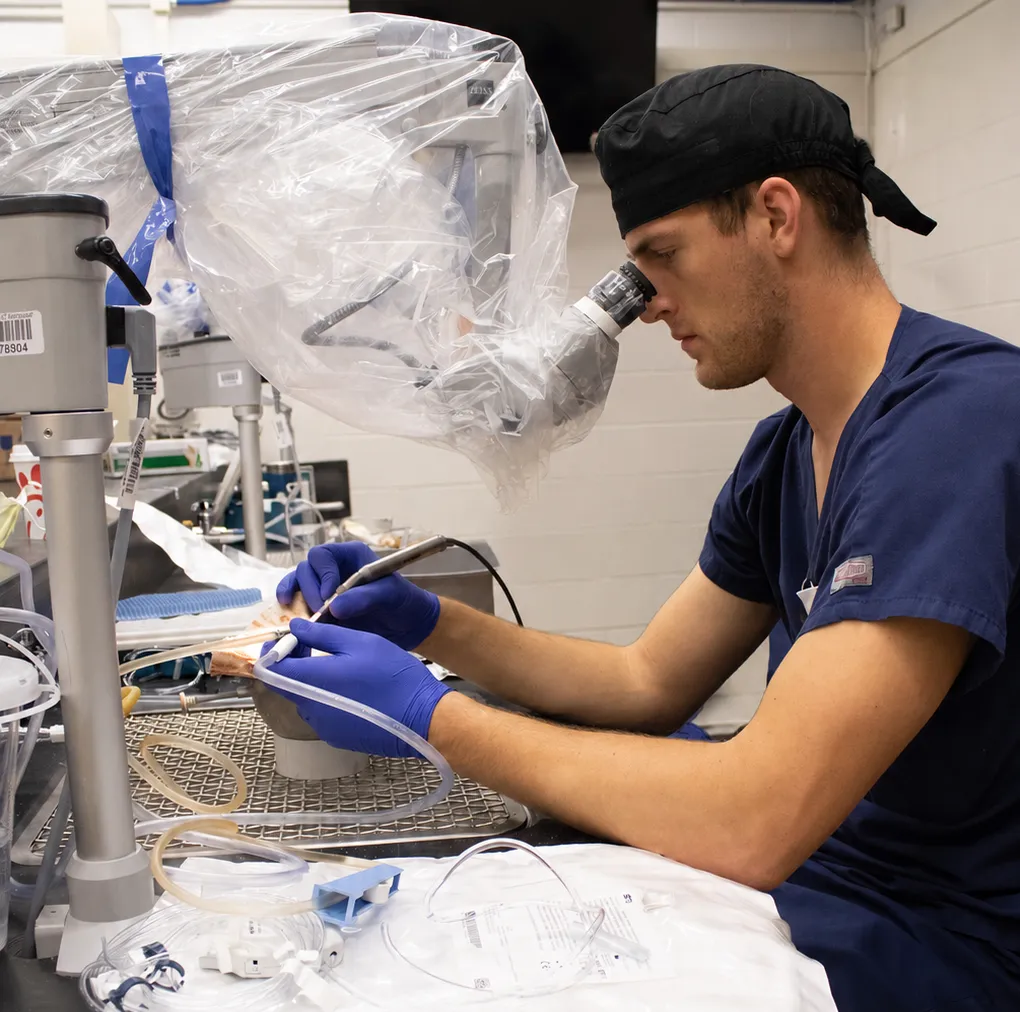
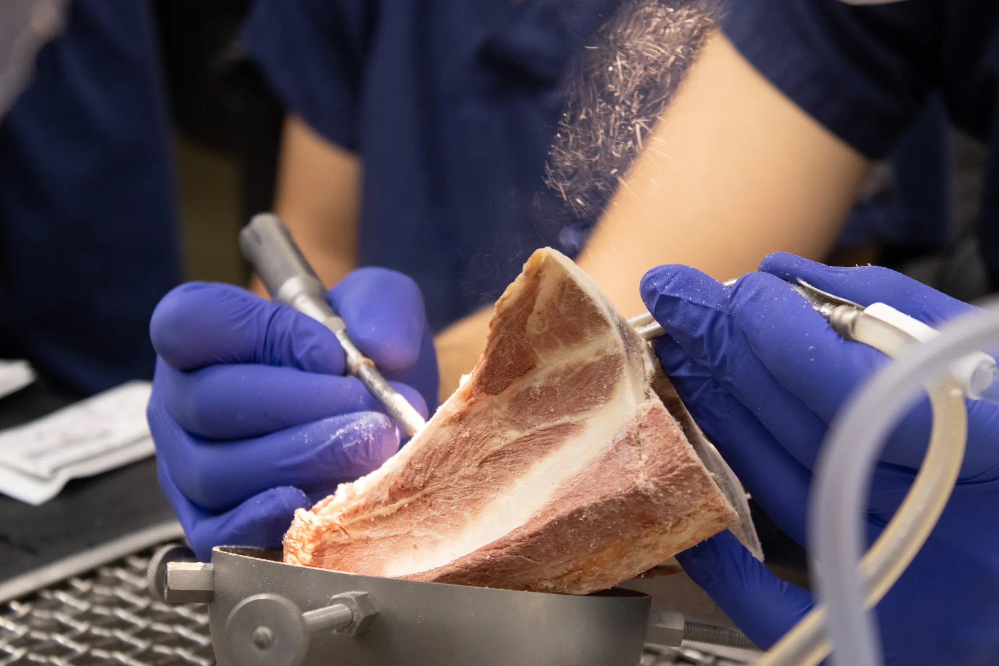
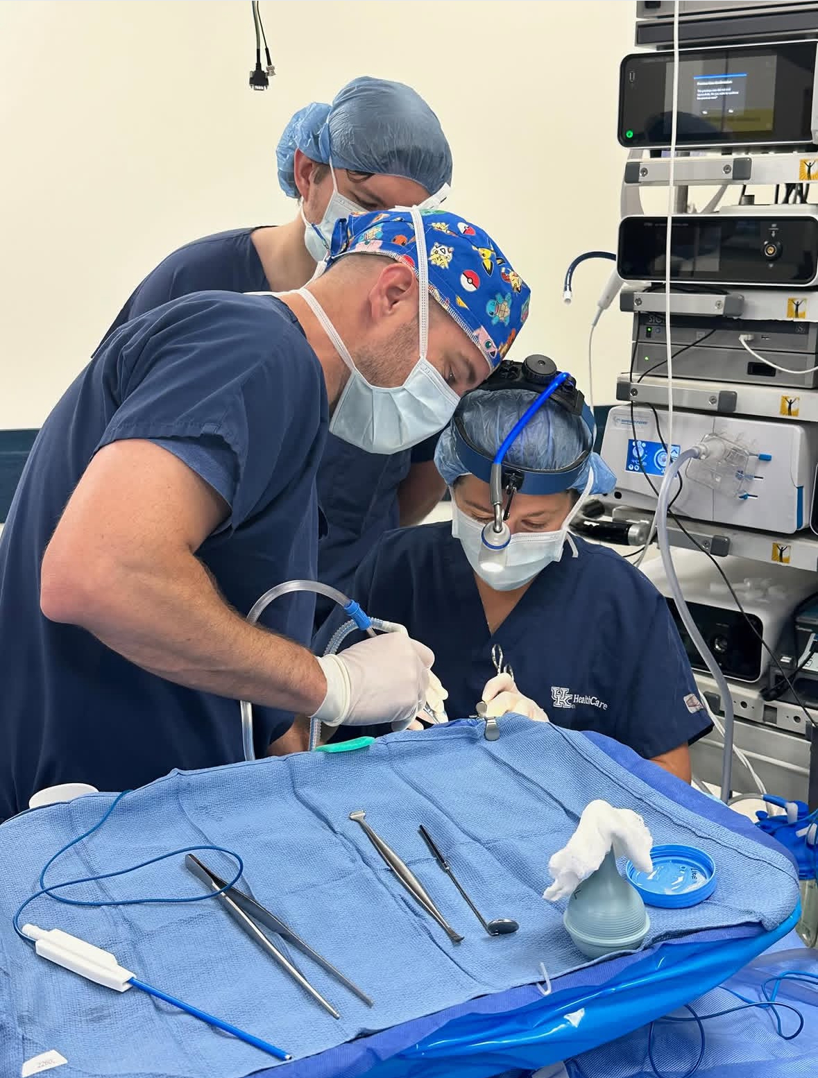
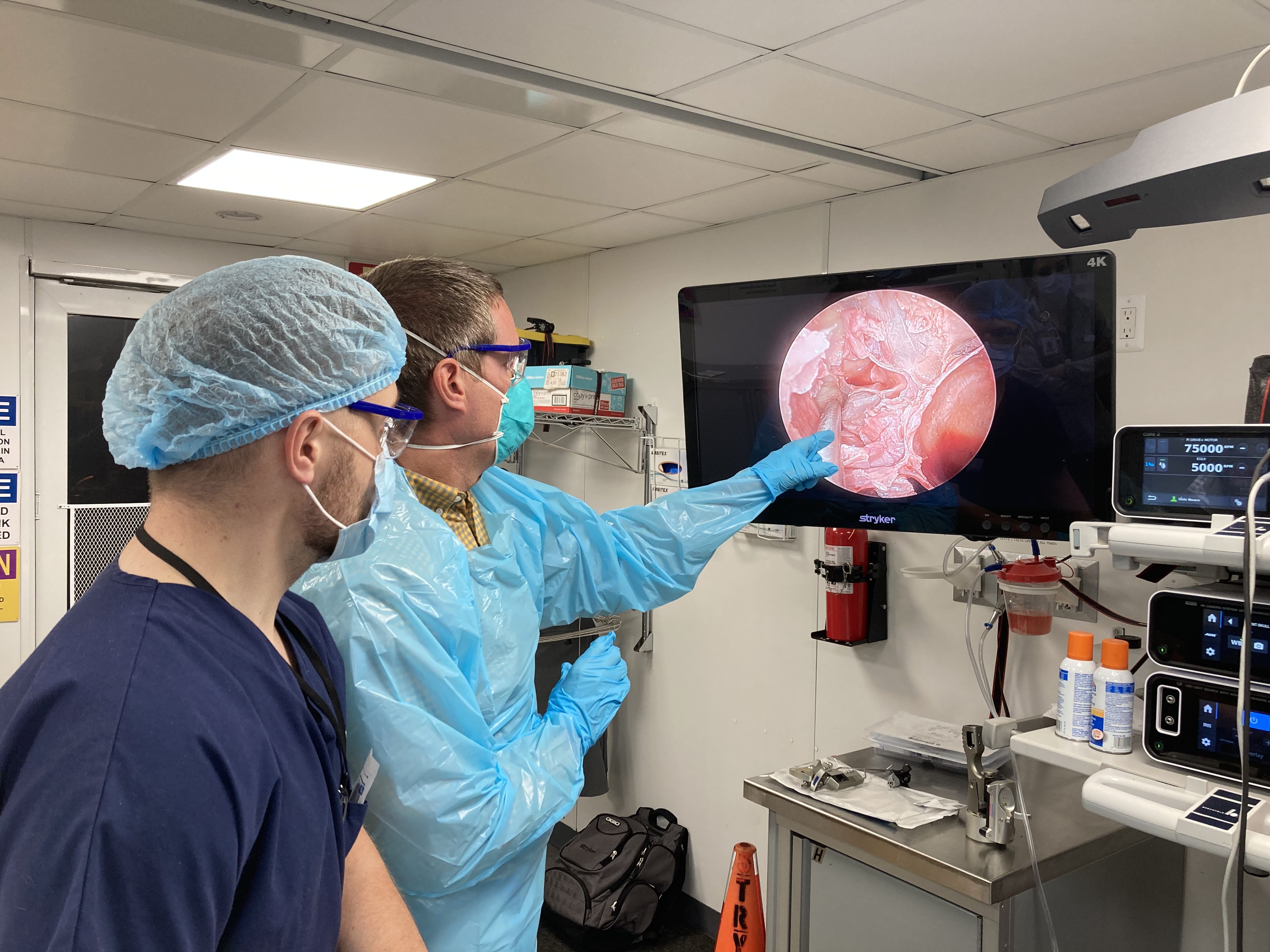
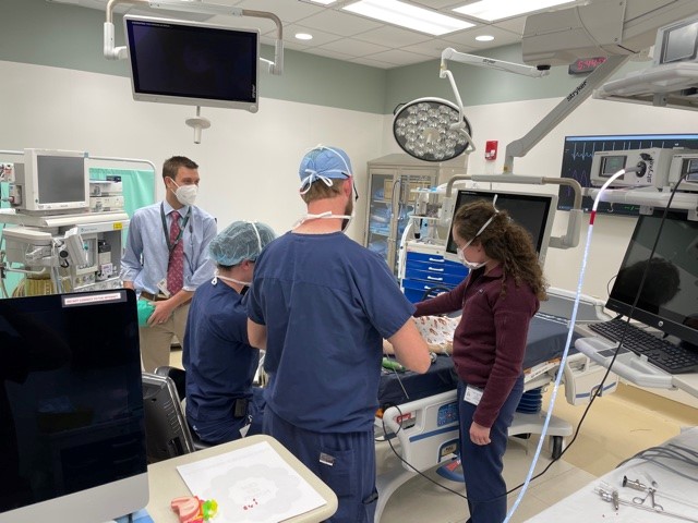
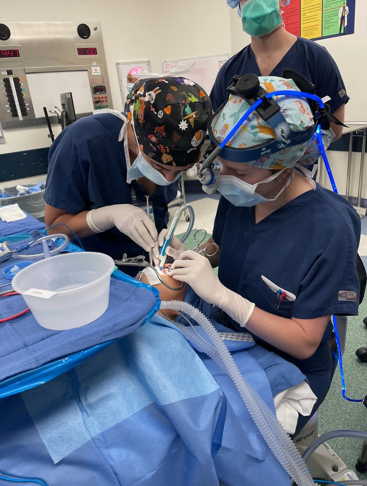
To support this approach, simulation experiences are incorporated throughout the year and paired closely with Resident Teaching Series (RTS) topics whenever possible. These experiences include temporal bone dissection during neurotology training, sinus dissection labs within rhinology education, allergy injection training, maxillofacial plating for facial plastic and reconstructive surgery, and pediatric airway foreign body removal simulations in pediatric otolaryngology. Through these opportunities, learners are able to build technical skills, confidence, and clinical decision-making in a safe and supportive environment.

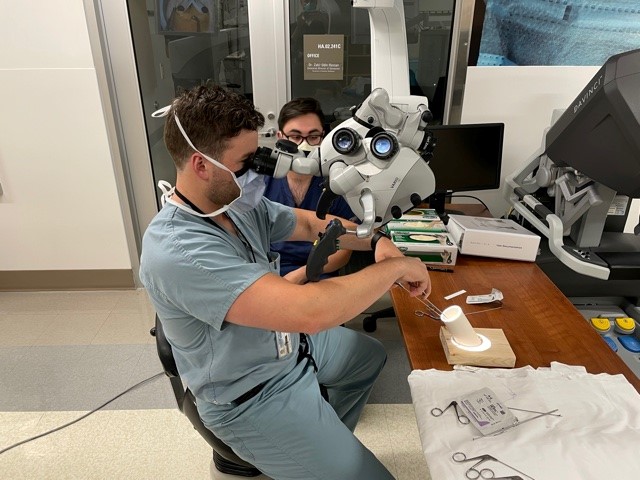
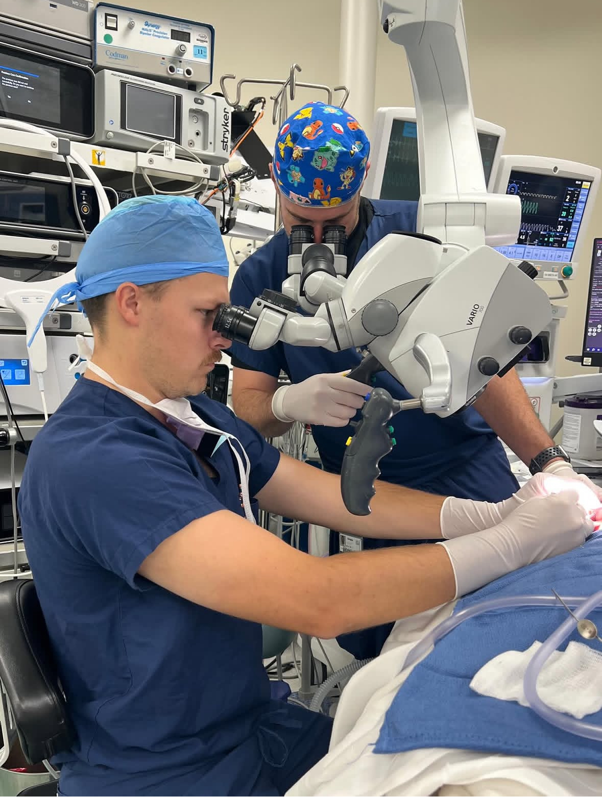
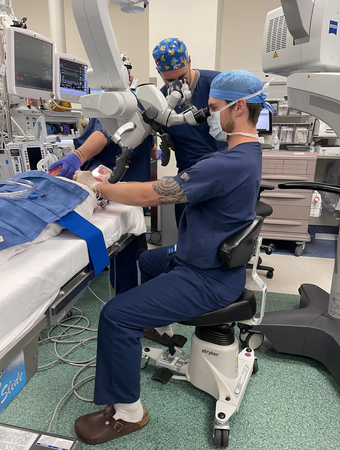
In the newly renovated Jason B. Diamond Temporal Bone Lab, residents participate in advanced cochlear implant drilling simulations.