MRI Used to Study Possible Therapies for Adult Congenital Heart Disease
Studies show that adults who received corrective surgery for the most common serious form of congenital heart disease as infants are susceptible to heart failure in adulthood.
Researchers at the University of Kentucky are using magnetic resonance imaging (MRI) technology to better understand the cause of heart failure in these patients, with the goal of eventually developing new therapies to reduce mortality. The team, led by University of Kentucky professor Dr. Brandon Fornwalt, recently published their findings in an article appearing in the European Heart Journal titled, "Patients with Repaired Tetralogy of Fallot Suffer From Intra- and Inter-Ventricular Cardiac Dyssynchrony: A Cardiac Magnetic Resonance Study."
The most common type of birth defect, congenital heart disease describes a problem with the structure of the heart that is present at birth. Many patients who are born with congenital heart disease receive surgeries as infants that will allow them to grow into adulthood. Adults living with congenital heart disease are now a growing population with complex medical problems. About two million adults in the United States are living with congenital heart disease, which accounts for more than double the years of life lost compared with that of all forms of childhood cancer combined. Hospital costs for congenital heart disease have quadrupled in the past seven years.
Tetralogy of Fallot is the most common serious form of congenital heart disease. The mortality rate of patients with tetralogy of Fallot triples 25 years after the initial surgery, and heart failure accounts for two-thirds of those deaths. In most cases, the surgical repair of tetralology of Fallot creates a disruption in the electrical system of the heart known as a right bundle branch block. This right bundle branch block could contribute to the development of an uncoordinated, or “dys-sychronous,” contraction in the heart. Dyssynchrony may play a role in the development of heart failure in these patients, and could potentially be treated with a pacemaker. However, significant research needs to will be required to investigate the pacemaker as a potential treatment option.
The University of Kentucky collaborated with several academic institutions to determine whether patients with repaired tetralogy of Fallot suffer from cardiac dyssynchrony. Through advanced cardiac MRI technology available at UK, a research team quantified the location and extent of dyssychrony in the hearts of healthy control subjects and patients with repaired tetralogy of Fallot. By observing patterns of contraction in the hearts using MRI, the researchers were able to identify dyssychrony in specific regions of the heart in the patients with tetralogy of Fallot. Their goal is to ultimately understand whether this dyssynchrony leads to cardiac failure long-term, and whether a pacemaker could potentially be used as a way to reverse the dyssynchrony and ultimately improve mortality in patients with tetralogy of Fallot.
"The goal of our research is to use imaging to change the way we practice medicine and ultimately improve lives," said Fornwalt, an assistant professor and researcher in the departments of pediatrics, biomedical engineering, electrical and computer engineering, physiology and cardiology at UK. "This study represents a small step in that direction, but we have lots more work to do to truly understand whether patients with tetralogy of Fallot might benefit from a pacemaker long-term."
This study was supported by the National Institutes of Health through the Director’s Early Independence Award Program and a grant to the University of Kentucky Center for Clinical and Translational Science from the National Center for Research Resources and National Center for Advancing Translational Sciences.
MEDIA CONTACT: Elizabeth Adams, elizabethadams@uky.edu

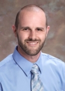
brandon_fornwalt_0.jpg