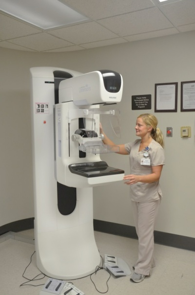
Mammography
Mammography uses ionizing radiation (x-rays) to create images of the human breast. Mammography can detect breast cancer.
Facilities, Technical Capabilities, and Limitations
- There are four mammography units available for human research.
- All units are capable of tomosynthesis (“image slices”).
- All units produce high-quality digital images.
- Images are viewable on Hologic SecurView workstations and archived on Enterprise-wide McKesson PACS; CDs/DVDs can be created as needed.
- Radiology reports are created in MagView, viewable through SCM/AEHR, and can be printed as needed.
| Units | Location | Type |
|---|---|---|
| 4 | Second Floor, Whitney-Hendrickson Building (Pavilion WH) | Hologic Selenia Dimensions (Digital Breast Tomosynthesis and Intelligent 2D Technology) |
| 1 | Second Floor, Whitney-Hendrickson Building (Pavilion WH) | Hologic Affirm Stereotactic Biopsy System (Upright) |
Hours of Operation
- Monday – Friday, 7:30 a.m. – 4:30 p.m.
- Other days/times by arrangement.
Personnel Resources
- American Board of Radiology-certified, subspecialized Radiology Physicians (MDs).
- American Board of Radiology-certified Medical Physicist (PhD).
- American Registry of Radiologic Technologists (ARRT)-certified Radiology Technologists with subspecialty certification in Mammography.
Costs
- Please discuss project and all applicable costs with Radiology Senior Research Coordinator prior to IRB and/or grant submission or renewal.
- Radiology costs include the technologist’s time to prepare and image the patient/human research subject according to protocol, process the image data, and archive the images to a CD/DVD if required by the investigator or study sponsor.
- Supply costs might be incurred, e.g., CDs/DVDs.
- Professional services by radiology physicians and/or medical physicists are negotiable.
Image